HNY Policy and Pathway Repository - York
Chronic unexplained breathlessness
Definition/Description
This document has been written to introduce the concept of a pathway that aims to address the multifactorial nature of chronic breathlessness in our locality. It amalgamates the existing cardiac and respiratory pathways to offer a holistic diagnostic process and breaks the current siloed, repetitive, and inefficient approach. The benefits for clinicians and patients are reduced time to diagnosis, appropriate specialist involvement, direct access to specialist expertise in respiratory and cardiac science for primary care providers and more decisive actions based on physiological evidence.
The proposal hopes to lay out the pathway's scope, the patient population it targets, investigations and the outcomes it hopes to achieve.
Red Flag Symptoms
CONSIDER RED FLAGS
Respiratory
- Haemoptysis, suggestion of Cancer
- Confusion, unable to speak in sentence and/or stridor.
- Unexplained reduction in SpO2 2000 ng/L
- Disproportionate respiratory rate
- Rapidly progressing breathlessness and reduction in functional capacity (within 4 weeks):
- Rapidly Progressing Muscle weakness/Dysphagia
Cardiac
- Persistent chest pain.
- BNP >2000 ng/L
- Significant ECG abnormalities (e.g. High degree AV block)
- Loss of Consciousness or blackout
Guidelines on Management
SCOPE
The patient group targeted are patients who present with ongoing breathlessness with no known cause. They should be patients with no known cardiac or respiratory pathology or already under specialist care.
The pathway is open to receiving referrals from GPs and all clinical primary care staff (e.g., ACP, Paramedics Practice Nurses) in the future.
This service is clinical scientist led with input/oversight from specialist consultants (i.e., respiratory, cardiac consultants) where required.
The key focus points of the pathway are:
- Identifying the most appropriate investigations to perform initially. Escalation of additional advanced investigations if required/clinically appropriate.
- Gather relevant diagnostic information for a more complete clinical picture for specialist review/decision making.
- Notification of GPs with results, management, advice, and outcomes.
REQUIREMENTS & PRE-CHECKS
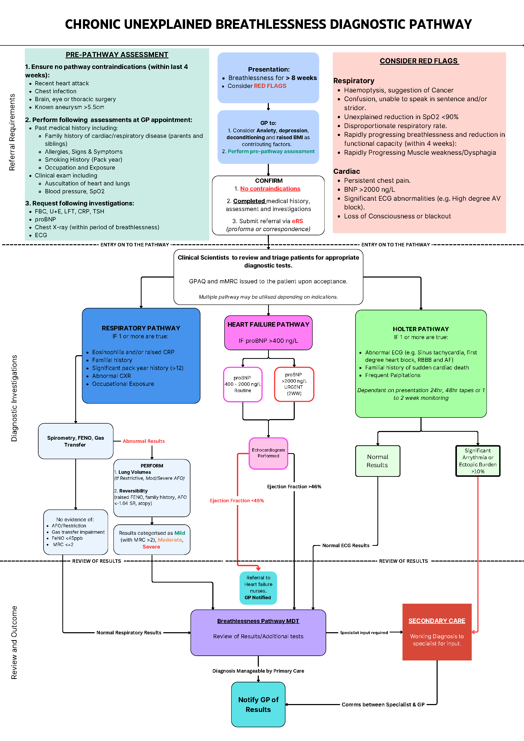
Patients referred to this pathway must have reported symptoms of breathlessness lasting more than 8 weeks. During clinical assessment, other causes of breathlessness (e.g., Anxiety, depression, deconditioning, increase BMI) should be considered prior to referral.
Red flags have been included as a safety net. Patients presenting with red flags may require urgent medical attention or intervention from specialist teams and existing pathways. Referrers should ensure they consider red flag features prior to referral. Existing urgent pathways such as 2WW Cancer, rapid chest pain clinic and urgent heart failure are already in place for high-risk patients.
There are initial investigations and history required to effectively triage patients for an appropriate investigation. It is important that the information given is complete and accurate to allow clinical scientists to identify the most suitable investigation(s) that should be performed. Below (in Table 1) is a list of the checks and the rationale for inclusion within the pre-check requirements for the pathway.
Once the above checks are completed (entered via referral proforma or correspondence), The referrer should ensure the following:
- No Red flags present.
- No contraindication met.
- Considered if the patient is suitable for other pathways following assessment and pre-checks.
Table 1. PRE-CHECKS to be performed by GP prior to referral to the pathway. |
|
Check |
Reason |
Past Medical History |
List any existing diagnosis (e.g., diabetes) and current medication. |
FBC, LFT, U+E, CRP, TSH |
Biomarkers used as inclusionary or exclusionary indicators. |
proBNP |
Biomarker for Heart failure |
Chest Xray |
To aid in determining anatomical abnormalities that may cause breathlessness. |
12 Lead ECG |
Identify any arrhythmia for further cardiac investigations |
Pack Year History |
To identify the extent of exposure. |
Familial History |
Identifying potential hereditary conditions |
History of palpitations |
Indication for cardiac investigation |
Auscultation of Heart and Lungs |
Normal or abnormal sounds (e.g., murmur, crackles) |
Occupational History |
Exposure to known hazardous substances from the work environment may cause breathlessness. |
Once checks are complete, if there is an obvious cause of breathlessness, the referrer should directly refer for relevant investigation (e.g. Heart failure, Asthma) as per NICE Guidance. Although, if the cause of the breathlessness is still not clear, multifactorial or complex following assessment, the patient should be referred to the breathlessness pathway.
Referral Criteria/Information
ENTRY INTO THE PATHWAY
Once the referral has been submitted via eRS, cardiac and respiratory Clinical Scientists shall triage the referral to identify the most appropriate investigation(s). Incomplete referrals will be rejected, as the lack of clinical information may lead to inefficient use of resources.
Some indications for respiratory investigations are raised biomarkers, familial history, significant pack year history, abnormal chest Xray (e.g. Fibrotic changes, hyperinflation, mucus plugging) and occupational exposure.
Heart failure pathway uses an inclusion criterion of a proBNP of >400ng/L as recommended by NICE.
Findings from the ECG, patient symptoms, and/or family history may require Holter monitoring (i.e., 24hrs, 48hrs)
In some cases, the patient may need to go down one or more diagnostic pathways based on their presentation (i.e., respiratory and heart failure). These would run concurrently to ensure results are available for analysis further down the pathway in a timely manner. There should be reasonable time given for this based on capacity and service demands.
Once the patient has been triaged and accepted onto the pathway, self-reported questionnaires are issued to the patient digitally, via correspondence or in person by the cardiorespiratory team. The questionnaires are:
- GPAQ (General Practice Activity Questionnaire): To gather activity level.
- MMRC (Modified Medical Research Council Questionnaire): Used to quantify dyspnoea.
DIAGNOSTIC INVESTIGATION
Patients are sent correspondence to attend the clinic for diagnostic testing. These investigations are performed by specialist Clinical Scientists with support from Practice Nurses and Healthcare Assistants.
Respiratory Pathway
The diagnostic tests performed are as follows:
- Spirometry.
- Gas Transfer.
- Lung Volumes (if there is a restriction or moderate/severe airflow obstruction present)
- Reversibility studies (Performed if the FEV1/FVC falls below –1.04 Z-score, history of atopy, persistent cough and wheeze).
Following these tests, patients are classified into normal, mild, moderate, or severe groups and reported breathlessness is reviewed. The referrer is notified of normal and abnormal results. Depending on the pathology found, it may be appropriate for patients to be managed in primary care (e.g., mild asthma or COPD). Although, more sinister pathologies may require follow-up in secondary care (e.g., interstitial lung disease). This decision would be made at an MDT with clinical scientists and respiratory consultants. There may be indication for further advanced diagnostic tests (e.g., Cardiopulmonary Exercise Test, bronchial provocation challenge etc.) to aid diagnosis if appropriate. The results from this shall be fed back to the referrer regardless of management in primary care or the requirement for secondary care intervention.
Heart Failure Pathway
The diagnostic tests performed are as follows:
- Transthoracic Echocardiogram
NICE state that a raised BNP >400 ng/L is an indication for heart failure. A proBNP > 2000 ng/L is significant and warrants urgent action. In both cases an echocardiogram shall be performed; a routine echocardiogram with a BNP 400-2000ng/L and urgent echocardiogram within 2 weeks if the BNP is >2000ng/L. Scans shall be performed by a British Society of Echocardiography (BSE) accredited sonographer.
The metric used to define significance in results is ejection fraction (EF). EF is a metric for systolic function. An ejection fraction (EF) of >40% is considered normal and <40% is considered impaired. An EF of 40% and below will trigger a referral to the heart failure nurses for treatment and management. If a patient’s EF is >40%, this is considered a normal result, and the GP shall be notified.
EF is not observed in isolation, other measurements of cardiac function are also assessed, including valvular impairment, chamber size and probability of pulmonary hypertension.
Holter Monitor Pathway
There are a few options for monitoring depending on presentation and frequency of symptoms, they are as follows:
- 24 and 48hr tapes
- 1- or 2-week monitoring
In conjunction with fitting the monitor, the patients are given a diary sheet to record activity and any symptoms they may have. When the monitors are returned, they will be analysed by specialist cardiac physiologists/scientists. The results will then be communicated back to the GP or escalated to secondary care if needed based on the findings.
REVIEW AND OUTCOMES
Following investigation(s), results are reviewed and electronically transferred to referrers. The report generated will summarise the investigations carried out, results obtained and an interpretation. Any additional advice on management, treatment and/or intervention by secondary care would be stated. The various pathways have different outcomes, which are detailed below.
RESPIRATORY
Following review at the MDT and any additional tests that may be requested. If a diagnosis is reached and where appropriate, GPs shall be notified with corresponding guidance and advice on management. Below are some examples of common and complex cases that may be managed in primary care or escalated to secondary care.
- Primary Care Management (As per NICE Guidance)
- Mild or moderate Asthma
- Mild or moderate COPD
- Secondary Care Intervention
- Severe/Uncontrolled Asthma
- Evidence of Interstitial Lung Disease (e.g., connective tissue disease, fibrosis)
- Evidence of pulmonary hypertension
- Severe COPD that may require lung volume reduction surgery.
- Dysfunctional Breathing
- Post COVID Syndrome
HEART FAILURE
EF is measured at <40%, patients are referred to heart failure nurses for treatment and management. The referrer shall be notified of this. The cardiac consultants shall also be notified if required.
If the echocardiogram shows other incidental findings (e.g., valvular dysfunction, right heart dilation) the referrer shall be notified with lifestyle and national guidance for management. The patient shall be escalated to cardiology for management if escalatory criteria is met.
HOLTER PATHWAY (PALPS Pathway)
After analysis, any significantly abnormal ECGs found would be escalated to cardiology. These abnormal rhythms include:
- Ventricular arrhythmia ≥30 beats.
- Complete heart block (diurnal/nocturnal) and high grade/2:1 AV block (diurnal or symptomatic)
- Frequent symptomatic second-degree AV block Type II (Mobitz II)
- Symptomatic or diurnal pauses of >3 seconds with a history of syncope.
- Symptomatic or diurnal pauses > 4 seconds in atrial fibrillation patients.
- Significant nocturnal pauses (consider the patient’s heart rate/rhythm and history).
- Atrial fibrillation with symptomatic fast ventricular response >150 bpm or an average HR>120bpm
- Ectopic burden >10%
The GP and the patient shall be notified of normal results with advice for patient management and prevention of symptoms.
ON WARD REFERRAL
If it is deemed necessary to seek further advice and management beyond the cardiorespiratory remit, on ward referral to other specialist within secondary care shall be place (e.g. ENT, Pulmonary Rehab, Oxygen Service etc). The referring GP shall be notified when this has occurred.
Future Developments
It is important to note that the pathway and its various elements are subject to change, given additional information, changes in guidance and/or service improvements via audits and evaluation. Any changes shall be communicated via email correspondence to primary care providers and stakeholders.
Additional Resources & Reference
Information sourced from: Y&STH Chronic Unexplained Breathlessness Diagnostic Pathway
Chronic Unexplained Breathlessness Diagnostic Pathway Flowchart (also below)
Appendix
At risk Occupations: COPD causes - occupations and substances (hse.gov.uk)
GP Activity Questionnaire: General Practice Physical Activity Questionnaire (publishing.service.gov.uk)
Modified MRC: Modified MRC Scale
Associated Policies
Specialties
Places covered by
- Vale of York
Hospital Trusts
York and Scarborough Teaching Hospitals